The cardiac arrhythmia and pacemaker unit provides diagnostic studies for the treatment of fast heart rhythms (tachycardia) and slow heart rhythms (bradycardias).
We also offer advanced studies and technology to diagnose your cardiac problem in relation to the electrical system of your heart and in addition, we offer healing through electrophysiological studies and ablation therapy.
Similarly, we offer the services of implantation of pacemakers, automatic defibrillators, and cardiac synchronizers through our electrophysiology laboratory and we perform programming of your pacemaker to perform your normal life.
Services we perform:
- Diagnosis and Treatment of Arrhythmias.
- Unicameral and Bicameral Definitive Pacemakers.
- Automatic Implantable Defibrillator.
- Cardiac Resynchronizer (Tricameral Pacemaker).
- Electrophysiological Study and Radiofrequency Ablation.
- Electrophysiological Study and Cryoablation.
- Syncope.
What is an arrhythmia?
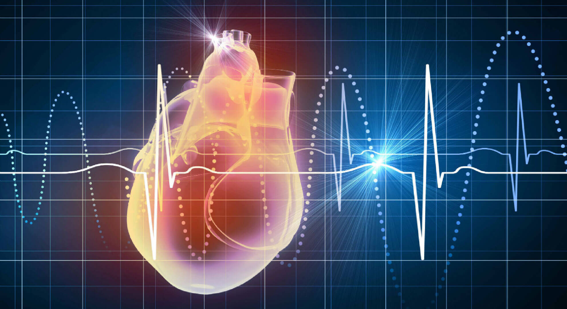
Arrhythmias are problems with the heart rate or rhythm of the heartbeat. During an arrhythmia the heart may beat too fast (tachycardia), too slow (bradycardia) or irregularly.
Most arrhythmias are harmless, but some can be serious and even life-threatening. When the heart beats too fast, too slow or irregularly, it may not be able to pump enough blood to the rest of the body. Lack of blood circulation can cause damage to the brain, heart and other organs.
Your heart is able to pump blood without working harder than necessary. To help make this happen, your heart has an electrical system that ensures that it contracts (pumps blood) in an orderly fashion. Arrhythmias are caused by problems with the heart's electrical conduction system.
Several factors can cause heartbeat irregularities. In some people, arrhythmias are a congenital defect, that is, they are born with this problem. Some diseases such as myocardial infarction, high blood pressure, heart growth, among others can contribute to arrhythmias. In addition, stress, caffeine, tobacco, alcohol, energy drinks, drugs and some over-the-counter cough and cold medicines can affect the natural rhythm of the heartbeat.
Major risk factors
Arrhythmias are more common in people who have diseases or health problems that weaken the heart, such as the following:
- Myocardial infarction.
- Heart failure or cardiomyopathy (heart growth), which weakens the heart and alters the way electrical impulses travel through the heart.
- Heart tissue that is too thick or stiff, or that has not formed normally (scar or hypertrophy).
- Heart valves that leak blood or are thinned, which overwork the heart and can cause heart failure.
- Congenital heart abnormalities (problems present at birth) that affect the structure or function of the heart. Arrhythmias may also be found in patients with apparently normal hearts.
Other health problems that can also increase the risk of arrhythmias include the following:
- High blood pressure.
- Infections that injure the heart muscle or the pericardium (sac surrounding the heart).
- Diabetes, which increases the risk of high blood pressure or heart attacks.
- Sleep apnea (shallow or stopped breathing during sleep), mainly in very obese patients.
- Hypothyroidism or hyperthyroidism (excess or lack of thyroid hormone in the body).
Many arrhythmias do not cause signs or symptoms so we call them silent.
When there are signs or symptoms, the most frequent are the following:
Signs and symptoms that may indicate seriousness are:
When there are signs or symptoms, the most frequent are the following:
- Palpitations (sensation of the heart beating very fast, very hard or referred to as fluttering). Some patients call this "I've got tachycardia".
- Slow heartbeat (Bradycardia)
- Irregular heartbeat.
- Pauses between one heartbeat and the next.
Signs and symptoms that may indicate seriousness are:
- Anxiety.
- Weakness, dizziness and lightheadedness.
- Fainting or feeling like you are about to faint.
- Sweating.
- Shortness of breath.
- Chest pain
Arrhythmias can be diagnosed using the following techniques:
- The electrocardiogram is the most effective test to diagnose arrhythmia. It records the electrical currents produced by the heart and determines the type of arrhythmia and in many cases the specific location within the heart. It should be performed at the moment the arrhythmia is present.
- The Holter monitor provides a continuous reading of the heart rate and rhythm over a 24-hour period (or longer). The patient wears a recording device (the Holter monitor), which is connected to small metal disks called "electrodes" that are placed on the chest. With certain types of monitors, the patient can press a recording button to record the heart rhythm as soon as he or she has symptoms. Physicians can study the recorded recordings on a computer to determine the type of arrhythmia and some characteristics of the arrhythmia.
- It is also possible to know the heart rhythm with monitoring systems of longer duration such as Implantable Holter, which are small devices that are placed under the skin in the fat located in the thorax with a wound of approximately 1 cm and allows to evaluate the heart rhythm for approximately 3 years.
- Electrophysiological studies are usually performed in a cardiac catheterization laboratory. A long, thin tube called a catheter is inserted into a vein or artery in the groin until it reaches the heart. The catheter captures the heart's electrical impulses, allowing a map of its electrical conduction system (electrocardiogram taken from inside the heart) to be obtained. This makes it possible to determine the type of arrhythmia and where it originates. During the study, physicians may administer controlled electrical impulses to see how the heart reacts, and certain medications may be administered to determine which ones eliminate the arrhythmia. When the electrical conduction pathways that produce the arrhythmia are discovered, electromagnetic waves can be sent through the catheter to destroy them by means of heat much like a microwave oven.
How is arrhythmia treated?
When treated in the emergency department, often the first measure to treat the arrhythmia is the administration of antiarrhythmic drugs, such as adenosine, digitalis, beta-blockers and calcium channel blockers. Other treatments include transcatheter interventions, implantable devices and surgery (in extreme cases).
- Ventricular tachycardia and ventricular fibrillation can be treated by placement of an implantable cardioverter defibrillator (ICD), a device that delivers rapid electrical stimulation of the heart or an intense electrical shock, to restore the heart's normal rhythm.
- In some cases of low heart rate, an electronic pacemaker is used. Smaller than a matchbox, the electronic pacemaker is surgically implanted near the collarbone, the bone in the shoulder. The pacemaker's batteries supply the electrical energy that acts like the heart's natural pacemaker.
- Radiofrequency ablation:This is a procedure in which a catheter and a device are used to map the electrical conduction pathways of the heart. Local anesthesia is used, a catheter is introduced through a vein in the groin until it reaches the heart. Using high-frequency electromagnetic waves, physicians can destroy (radiofrequency ablation) the conduction pathways responsible for the arrhythmia with a very high percentage chance of cure.
Remember: Arrhythmias must be treated by Cardiologists with a specialty in Cardiac Electrophysiology who will recommend the best treatment option with medication or offer you a cure with an Electrophysiological Study and Radiofrequency Ablation or placement of a definitive pacemaker with special functions according to your needs. Do not hesitate to visit the Arrhythmia and Pacemaker Unit.